Description
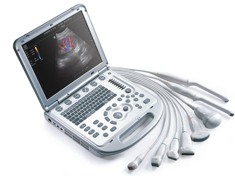
EDAN DUS-60 Portable B/W Ultrasound
Standard Configuration:
12.1 inch LCD monitor
Two standard transducer connectors
8-segment TGC control
Phased Inversion Harmonic Compound imaging
Multi-Beam-Forming
Synthetic aperture
IP(Image Processing) function
256-frame cine loop memory
504MB image storage capacity
Two USB ports
Pseudo color
Measurement&Calculation software package
Flash Disk (Netac, 4G, USB2.0 Protocol)
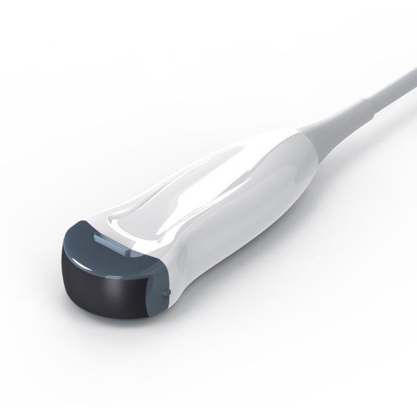
Transducer: C361-2 Convex
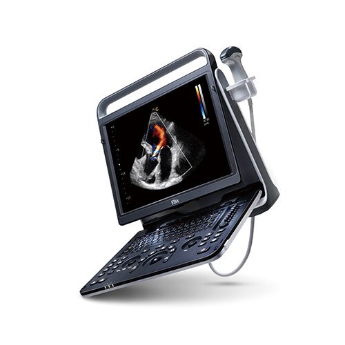
Compact, Lightweight Design
12.1″ TFT-LCD monitor
Two active and exchangeable connectors
Backlit, easy-to-use control panel
Multiple peripheral ports
Innovative Technologies
PW Doppler supplies more hemodynamics information
Five-frequency transducers increase versatility
Phase Inversion Harmonic Imaging provides best-in-class image quality
Practical tools enhance efficiency
Intelligent 8-segment TGC for precise adjustment
Multi-format data transfer via USB and DICOM
Multiple-pseudo-color options enhance image presentation
User-friendly Workflow
One touch image optimization via smart image processing key
User defined settings for personalized use
Two hours of battery-powered operation
Various Clinical Applications
Abdomen, Obstetrics, Gynecology, Endovaginal, Small Parts, Muculoskeletal, Vascular, Urology, Endorectal, Pediatrics
The DUS 60 provides outstanding performance for general imaging.
1.System Overview 1.1.
Application – Abdomen – Gynecology – Obstetrics – Cardiology – Small parts – MSK – Urology – Vascular – Pediatric
1.2.Transducer Types – Convex array – Linear array – Endocavity curved array – Endocavity linear array 1.3.Imaging Modes – B-mode – M-mode – Pulsed Wave Doppler
1.4.Imaging Technique & Function – Harmonic Imaging – Multi-Beam-Forming – Speckle Reduction Imaging (eSRI) – Synthetic Receiving Aperture – Pan Zoom – Needle Guide
1.5.Display Modes – B – B+B – 4B – M – B+M – B+PW
1.6. System Language Support – Software : English, Chinese, German, French, Italian, Russian, Spanish, Polish, Portuguese – Keyboard input: English, Chinese, Russian – Control panel overlay: English, Chinese – User Manual: English, Chinese
1.7. Standard Features – USB Disk (only for international marketing) – Probe cable holder – Dustproof cloth 1.8. Optional Features – Transducers – Printers – Battery (only for international marketing) – Hard disk – Needle Guide Bracket Kit – Mobile trolley – Footswitch – Hand carried bag – Luxury Hand carried bag – DICOM 3.0 2.
2.0 Physical Specification
2.1. System Architecture
– Physical Channels: 24
– System dynamic range: 0-158dB
– Beam forming: Dual beam
– Memory: 504MB
– Hard drive: 320GB(optional)
– Operation System: ucos
2.2. Dimension and weight
– Height: 32cm
– Width: 22cm
– Depth: 33cm
– Weight: 7.1kg 2.3. Monitor
– 12.1’’ TFT-LCD monitor
– Resolution: 1024 x 768
– Imaging field size: 640*512
– Video out size: 1024 * 680
– Capture size: BMP, JPG, AVI, DCM 1024 * 768; FRM, CIN 640 * 512
– View angle: Up 80°, Down 80°, Left 80°, Right 80°
– Brightness, Contrast and Color Temp adjustable
– Built-in stereo speaker
2.4. Transducer port and holder
– 2 active ports – 2holders
– 1 Coupling holder
2.5. Electrical Power
– Voltage: 110V-240VAC
– Frequency: 50/60 Hz
– Power: 74.4w 2.6. Battery
– Rechargeable lithium ion battery
– Capacity: 5000mAh
– Removable – Approximately 60 minutes of typical ultrasound exam use
– Max charging time:8 hours
2.7. Environmental operating requirements
– Ambient temperature: 5° to 40°C
– Relative Humidity: 25%~80% (no condensation)
– Atmospheric pressure: 86kPa-106kPa 2.8. Environmental storage requirements
– Ambient temperature: -20° to 55°C
– Relative Humidity: 25%~93% (no condensation)
Atmospheric pressure: 70kPa-106kPa
3.0 User Interface
3.1. Control Panel
– Interactive back-lighting
– Hard Keys provides tactile feedback
– Physical trackball
– 8 segment TGC sliders
– Physical keyboard
3.2. System boot-up
– Boot up from complete shut-down in about 30 sec
– Shut-down in about3 sec
– Recovery from screen saver in about 3 sec
3.3. Comments
– Arrow
– Block move and delete for separate blocks of text
– Support physical keyboard for text input
– 150 comments in comments library
– 15 User customizable comments per a library
3.4. BodyMark
– Up to 130 Body Mark graphics in library
3.5. Screen Information
– EDAN logo
– Hospital name
– Date
– Time
– Patient ID
– Patient Name
– Transducermodel
– Preset name
– Mechanical index (MI)
– Thermal Index (TI)
– Imaging parameters
– Gray Scale bar
– Depth Scale *Not all the items are listedin here,please refer tothe User Manual. 4. Imaging
4.0 Parameters
4.1. B-Mode
– Zoom: 7 levels, x1.44,x1.96,x2.56,x4.0,x5.76, x9.0, x16.0 (available on live state); 3 levels: x1.78, x4.0, x16.0(available on freeze state)
– Depth: 1.9- 32.4cm
– Frequency: 2.0-15.0MHz(3 fundamental &2 harmonic frequencies)
– eSRI: 0-3
– Rejection: 0-7
– Scan Angle: 0-3
– Gain: 0-130dB, 2dB/step
– TGC: 8 segments
– Dynamic range: 30-150 dB, 4dB/step
– Scan Density: H/M/L
– Max Frame rate: 270 f/s – Map: 0-14
– Pseudo Color: 6 types
– Frame Persist: 0-7
– Focus position: 0-15
– Focus Number: 1-4
– GAC: 0-7
– IP: 0-7 – SRA: 0-1
– H Reverse: On/Off
– V Reverse: On/Off
– 90°rotation : 0/90/180/270
– B/W Invert: On/Off
– Display format: single(B),dual(B+B), Quad(4B):B,2B,4B
4.2. M-Mode
– Sweep speed: 0-3
– Line Average: 0-7
– Gray Map: 0-14
– Pseudo Color: 6 types
– Gain: 0-130dB, 2dB/step
– Frequency: 2.0-15.0MHz(3 fundamental frequencies)
– Dynamic range: 30-150 dB, step 4dB/step
– B/M Display: L/R, Full 4.3. Pulsed Wave Doppler
– Frequency: 2.5-6.5MHz, 2 levels – PRF: 13 levels – Gain: 0-130dB, 2dB/step
– Dynamic range: 30-150dB, 4dB/step
– Wall filter:0-3 – Sweep speed: 0-3
– Baseline: 0-6 – Correct Angle: 15° -165°, 1°/step
– Steer:0°,±10°(available on linear transducers)
– Invert: Up/Down – Volume: 0-7
– Pseudo color: 6 types
– Sample Volume: 16 levels, 0.5-20 mm
– D Rejection: 0-7
5. Cine Review and Post-Processing
5.1. Cine Review
– Frame by frame manual review
– Auto playback
– Start frame and end frame are selectable for cine loop review
– Maximum cine memory is up to: 256 frames for B mode 23s for M mode 10s for PW Doppler mode
5.2. Post-Processing
– B Mode: zoom, pseudo color, Gray map
– M Mode: pseudo color, Gray map
– PW: pseudo color, Gray map *Not available the stored images and clips in Review
– FRM and CINE file support measurement, comments, bodymark
6. Imaging Storage and Exam Database
6.1. Imaging Storage
– 504MB for data storage 504MB
– 320GB hard drive
– Storage up to approximately224static images as BMP format(memory); storage up to approximately 145635 static images as BMP format (hard drive)
– Maximum clip is up to: 256 frames for B mode 23s for M mode 10s for PW Doppler mode
6.2. File Management
– Support exam storage temprarily without patient information
– Support image files query – Support delete, rename image files
– Support review image files of current exam or prior exam
– Support store images as BMP, JPG, FRM, AVI, CIN or DCM format
– Support export images to a USB disk
7. Connectivity
– DICOM Storage: Verify SCP Static image store SCU DCM Removable media Manual-transfer on demand
– 2 USB Ports – Video out: VGA Video: PAL/NTSC S-video: PAL/NTSC
– Footswitch port – Remote port
– Ethernet
8. Preset
– Application: Abdomen Obstetric Gynecology Small Parts Urology Vascular Cardiac Pediatric – Presets Abdomen Abd Difficult Aorta Obstetric Gynecology Endovaginal Small Parts Urology Vascular Cardiac Pediatric
9. Peripheral& Accession
9.1. Printer
– Black/white Digital/Analog
Video printer SONY UP-X898MD MITSUBISH P93W_Z
– Color Analog video printer
SONY UP-25MD SONY UP-D25MD
– Graph/text printer
HP Laserjet Pro 400 M401d402D 403D
HP LaserJet 1510 HP Deskjet 1010
HP DeskJetlnk Advantage Ultra 2029
HP DeskJet 1112
9.2. Needle Guide Bracket
BGK-CR60 – Focus Depth:44mm – Angle:45°
BGK-LA43 – Focus Depth:26.5mm – Angle:43°
BGK-CR10UA – Focus Depth: 250mm – Angle: 3°
BGK-LA70 – Focus Depth:42.5mm – Angle:44°
BGK-MCR10 – Focus Depth:20mm – Angle:35°
BGK-EL40 – Focus Depth:23mm – Angle:0°
10. Measurement and Report
10.1. General Measurement
B Mode:
– Distance
– Cir/Area
– Volume: 2-Axis, 3-Axis, 3-Axis(LWH)
– Ratio
– Angle
– %Stenosis:Distance, Area
– Histogram
M Mode
– Distance
– Time
– Slope
– Heart Rate
Doppler –
Velocity
– Heart Rate
– Time
– Acceleration
– RI
– Auto: PS, ED, TAMAX, RI, PI, S/D
– Trace Direction: Above, below, Dual
– Trace Sensitivity+
– Trace Sensitivity
10.2. Application Measurement
Gynecology
B mode
– UT:Length, Width, Height, UT
– Endo
– OV-Vol(L/R): Length, Width, Height, OV-Vol
– FO(L/R, Number:4): Length, Width
– CX-L
– UT-L/CX-L
PW mode
– Velocity, L UT A, R UT A, L OV A, R OV A,
Trace Direction, Trace Sensitivity+, Trace Sensitivity-
OB
B mode-OB-1
– GS , CRL, BPD, HC, AC, FL, AFI, EFW
– FBP
– Growth Curve
– EDC B mode-OB-2
– TAD, APAD, CER, FTA, HUM, OFD, THD, NT – FBP – EDC
PW mode
– Velocity, FHR, Umb A, MCA, Fetal AO, Desc.AO, Placent A, Ductus V, Trace Direction, Trace Sensitivity+, Trace Sensitivity
M mode
– FHR, Time, Slope
Cardiac
M mode
– LV: TEICHHOLZ(LVIDd, LVIDs, ET, HR, EDV, EDS, SV, CO, EF, FS, SI, CI, MVCF, BSA),
CUBE(LVIDd, LVIDs, ET, HR, EDV, EDS, SV, CO, EF, FS, SI, CI, MVCF, BSA)
– Mitral Valve: EF Slope, ACV, A/E, Valve Volume
– Aorta: LAD/AOD, Valve Volume
– Heart Rate
– LVET: LVET, AVSA
– LVMW: LVPWd, IVSTd, LVIDd, LVMW
B mode
– RV
– LV: S-P Ellipse (LVLd, LVALd, LVLs, LVALs, EDV, ESV, SV, CO, EF, SI, CI, BSA), B-P Ellipse (LVALd, LVAMd, LVIDd, LVALs, LVAMs, LVIDs, EDV, ESV, SV, CO, EF, SI, CI, BSA), Bullet (LVAMd, LVLd, LVAMs, LVLs, EDV, ESV, SV, CO, EF, SI, CI, BSA), Mod. Simpson(LVAMd, LVLd, LVAPd, LVAMs, LVLs, LVAPs, EDV, ESV, SV, CO, EF, SI, CI, BSA)
– RV
– PA
Small Part B mode
– L.THY-V: Length, Width, Height, Volume
– R.THY-V: Length, Width, Height, Volume – Isthmus
Urology B mode
– BLV: Length, Width, Height, Volume
– RUV: Length, Width, Height, Volume
– Prostate Vol: Length, Width, Height, Volume, PPSA, PSAD
Pediatric B mode
– HIP(α, β)
Vascular PW mode
– Velocity, CCA, ICA, ECA, Vert A, Upper, Lower, Trace Direction, Trace Sensitivity+, Trace Sensitivity- *For more measurement information, please refer to the User Manual.
10.3. Report
– Worksheet
– Diagnostic
– Configure whether print image in report
– Export as PDF format
11. Transducers
C361-2
– Imaging Format: convex array
– Number of Elements: 80
– Convex Radius: 60 mm
– FOV:60°
– Bandwidth: 2.0-6.0MHz
– Fundamental Frequency: 2.5 MHz, 3.5MHz, 4.5MHz
– Harmonic Frequency: H5.0MHz, H5.4MHz
– Doppler Frequency: 2.5MHz, 3.0MHz
– Depth: 19-324mm
– Frame Rate(18cm, Full of FOV): max 103 f/s
– PW velocity: max 2.01m/s(±60°)
– Applications: Abdomen, OB, Gynecology, Urology
– Needle Guide Bracket: BGK-CR60(14G, 18G, 20G, 22G)
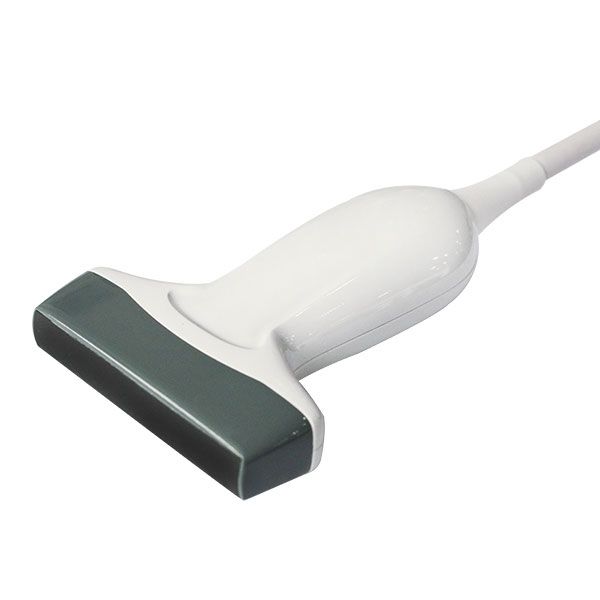
L743-2
– Imaging Format: general linear array
– Number of Elements: 96
– Footprint: 40 mm
– Bandwidth: 5.0-10.0MHz
– Fundamental Frequency: 6.5 MHz, 7.5MHz, 8.5MHz
– Harmonic Frequency: 9.0 MHz, 9.4MHz
– Doppler Frequency: 5.5MHz, 6.5MHz
– Depth: 19-117mm
– PW velocity : max 1.14m/s(±60°)
– Applications: SMP, Vascular
– Needle Guide Bracket: BGK-LA43(14G, 18G, 20G, 22G)
L761-2
– Imaging Format: general linear array
– Number of Elements: 80
– Footprint: 60mm
– Bandwidth: 5.0-10.0MHz
– Fundamental Frequency: 6.5 MHz, 7.5MHz, 8.5MHz
– Harmonic Frequency: 9.0 MHz, 9.4MHz
– Doppler Frequency: 5.5MHz, 6.5MHz
– Depth: 29-117mm
– PW velocity : max 1.14m/s(±60°)
– Applications: SMP, Vascular
– Needle Guide Bracket: BGK-LA70(14G, 18G, 20G, 22G)
E611-2
– Imaging Format: endocavity micro convex array
– Number of elements: 80
– Convex Radius: 10 mm
– FOV: 155°
– Bandwidth: 5.0-8.0MHz
– Fundamental Frequency: 5.5 MHz, 6.5MHz, 7.5MHz
– Harmonic Frequency: 9.0 MHz, 9.4MHz
– Doppler Frequency: 5.0MHz, 6.0MHz
– Depth: 19-157mm
– Frame Rate(10cm, Full of FOV): max168 f/s
– PW velocity: max 0.56m/s(±60°)
– Applications: OB, Gynecology, Urology
– Needle Guide Bracket: BGK-CR10UA(16G)
C611-2
– Imaging Format: micro convex array
– Number of elements: 80
– Convex Radius: 10 mm
– FOV: 128°
– Bandwidth: 5.0-8.0MHz
– Fundamental Frequency: 5.5 MHz, 6.5MHz, 7.5MHz
– Harmonic Frequency: 9.0 MHz, 9.4MHz
– Doppler Frequency: 5.0MHz, 6.0MHz
– Depth: 29-157mm
– Frame Rate(10cm, Full of FOV): max 168 f/s
– PW velocity : max 0.79m/s(±60°)
– Applications: Pediatric, Pediatric Cardiac
– Needle Guide Bracket: BGK-MCR10(14G, 18G, 20G, 22G)
E741-2
– Imaging Format: endocavity linear array
– Number of elements: 80
– Footprint: 40 mm
– Bandwidth: 5.0-10.0MHz
– Fundamental Frequency: 6.5 MHz, 7.5MHz, 8.5MHz
– Harmonic Frequency: 9.0 MHz, 9.4MHz
– Doppler Frequency: 5.5MHz, 6.5MHz
– Depth: 19-117mm
– PW velocity : max 0.69m/s(±60°)
– Applications: Urology
– Needle Guide Bracket: BGK-EL40(16G, 18G)
12. Regulatory approvals
– FDA Class II Device
– CE/MDD Class IIa
– IEC 60601-1: Medical Equipment Safety
– IEC 60601-1-2: Medical Device Electromagnetic Safety
– IEC 60601-2-37: Ultrasonic Medical Equipment Safety
– IEC 62133: Battery Safety
– IEC 62304: Medical Device Software Lifecycle Process
– IEC 62366: Medical Device Usability Engineering
– EN ISO 14971: Medical Device Risk Management
– ISO 10993: Medical Device Biocompatibility
– NEMA UD 2: Output Measurement for Diagnostic Ultrasound Equipment
Additional information
| Weight | 15 kg |
|---|
Related products
Chison EBit 60 PW Colour Ultrasound
$17,500.00 + GST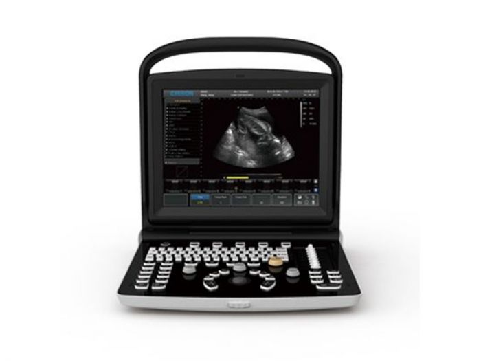
Chison ECO3 EXPERT B/W Portable Ultrasound with PW
$6,400.00 + GST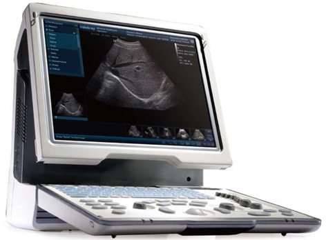
Chison MC3-V Micro Convex Probe for Sonobook
$3,800.00 + GST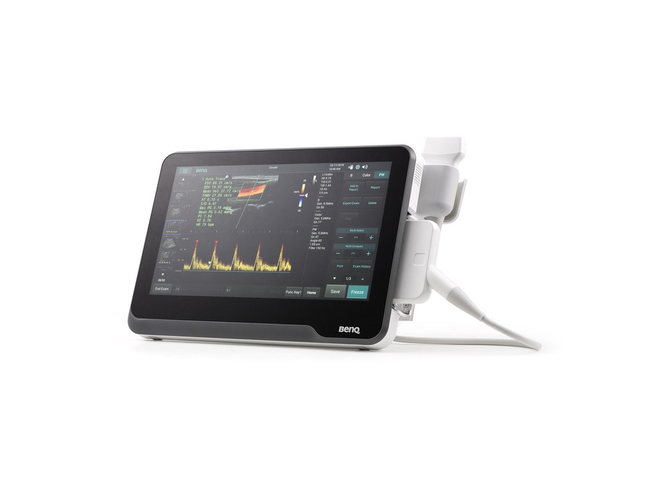
BENQ Medical T3300 Tablet POC Ultrasound
$19,400.00 + GST
Chison L8M5-V Linear Probe for Sonobook
$6,200.00 + GST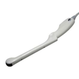
Chison E7W-V Intra Cavity Probe for Sonobook
$6,200.00 + GST